
Other Projects and Collaborations
SKIP TO:
Developing Shape Analysis Software (collaboration with Kitware)
Idiopathic Scoliosis in Children (collaboration with Dr. James Sanders)
Developing an Open-Source Automated Shape Analysis Pipeline for Bone
In collaboration with Kitware, Inc.
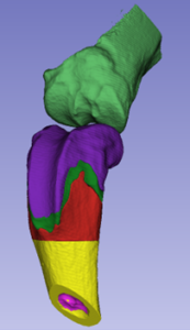
For any given bone, various surface features such as bumps, ridges, grooves, and knobs may not appear important at first glance, but this could not be farther from the truth. Each facet of a bone plays a vital role, be it an attachment point for muscle, a joint, contact surface, reinforcement to improve load-bearing function, or a combination of multiple roles. This holds true even below the bone surface. Interior features such as the size and shape of trabecular bone structures, marrow cavities, and growth plates can strongly influence various functions of bone. Many bone pathologies are likewise associated with changes to bone shape features. Therefore, shape analysis, a method for determining the size, shape, and position of distinct bone features, has great potential for versatile applications. In this project, we are testing a web-based, open-source bone shape analysis pipeline by quantifying differences between models for healthy and diseased bone created from 3D medical images. By comparing the statistical significance of these differences with standard metrics, we aim to show that our accessible pipeline is as effective as standard analyses performed with costly, specialized equipment.
Image: 3D model of a mouse knee segmented to identify anatomical shape features: tibia – light green, femur epiphysis – purple, femur growth plate – dark green, femur metaphysis – red, femur diaphysis – yellow, femur intracortical space – pink
Idiopathic Scoliosis in Children: Exploring Causes and Progression with 3D Image Analysis and Modeling
In collaboration with Dr. James Sanders,
UNC School of Medicine Department of Orthopaedics
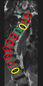
Idiopathic scoliosis in children presents as abnormal curvature of the spine that tends to initiate in late childhood or adolescence without any definite cause. Once a curve begins to form, it may worsen over time and in severe cases, mobility loss, pain, and restriction of lung volume may occur. Despite the cause remaining unknown, this project aims to investigate the role of the nucleus pulposus, a gel-like tissue typically in the center of the intervertebral disc. We are creating 3D models of scoliotic spines from MRI images to characterize deviation of the nucleus pulposus away from the center of the intervertebral disc and towards the convex side of the curve. This deviation may affect load transfer between vertebrae, which may impact vertebral growth as bone development and maintenance is strongly regulated by mechanical stimuli. In the second phase of this project, we will perform finite element analyses on the same 3D spine models to investigate patterns of stress and strain experienced by the vertebrae while supporting standing loads. Through this combination of techniques, we will determine whether deviation of the nucleus pulposus may be part of a positive feedback loop culminating in the progressive worsening of spinal curvature.
Image: Coronal view showing sideways spinal curvature. Nucleus pulposi are circled, and color coded to show the degree of in-plane deviation: green – not deviated, yellow – slightly deviated, red – highly deviated