
Previous Research
SKIP TO:
Examining Bone Vasculature using a Stroke Model (opens new page)
Aging and Exercise Effects on Bone and Tissue (collaboration with Dr. Maya Styner)
Barbed Surgical Sutures (collaboration with Dr. Martin King)
Musculoskeletal Aging (collaboration with Dr. Gregory Sawicki)
Tension Band Wiring Configurations for Stable Bone Fracture Fixation (collaboration with Dr. Simon Roe)
Meniscus for Improved Allograft Replacements (collaboration with Drs. Jeffrey Spang, Elizabeth Loboa, and Matthew Fisher)
Interplay of Aging and Exercise Effects on Whole Bone Bending and Tissue Elastic Modulus
In collaboration with Dr. Maya Styner, UNC School of Medicine
With age, bone tends to become more brittle, increasing the risk of sudden fracture under moderate to high loads. On the other hand, regular exercise with interspersed periods of rest stimulates bone formation, strengthening bone and reducing the likelihood of fracture. Unfortunately, habitual exercise is most common in youth and young adults, so the degree to which exercise benefits the elderly remains unclear. This project aims to determine the interaction effects of aging and exercise on the bending properties and tissue elastic modulus of mouse femora. Using a combined imaging and mechanical testing approach, we will not only examine the mechanical performance of bones in both exercising and non-exercising groups, but bone geometric and material properties as well to determine precisely how exercise may affect aging bone.
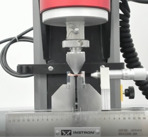
Mouse femur shown in a 3-point bending configuration for measuring bone bending stiffness
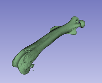
3D model created from micro-CT image of a mouse femur for analyzing bone geometry
Barbed Surgical Sutures: Evaluating the Performance of Barbed Surgical Sutures for Orthopaedic Applications
In collaboration with Dr. Martin King, College of Textiles, NC State
Barbed surgical sutures are knotless sutures approved by the US FDA in 2004 that have been used in wound closure in various fields, including dermatology, urology, and orthopaedics. However, many aspects of these sutures need to be explore further, including the material selection and performance of various polymers used for these sutures and the extension of usage to tissue repair surgeries. We are characterizing the anchoring performance and tissue engagement of barbed surgical sutures manufactured from various polymers and are evaluating the performance of these sutures in tendon repair using animal models.
Publications and Presentations:
- Huang Y, Cadet ER, King MW, Cole JH. Comparison of the mechanical properties and anchoring performance of polyvinylidene fluoride and polypropylene barbed sutures for tendon repair. Under Review.
- Cong H, Ruff GL, Cole JH, Tokarz DA, Roe SC, Liu W, King MW (2018) In vivo comparison of polydioxanone and polyhydroxyalkanoate barbed surgical sutures in a rat model. Society for Biomaterials Annual Meeting, Atlanta, GA, April 11-14, Podium #202.
- Cong H*, Ruff GL, Cole JH, King MW (2017) Hydrolytic degradation of polydioxanone barbed and non-barbed sutures. International Conference on Medical Textiles and International Forum on Biomedical Textile Materials, Shanghai, China, May 17-19, podium. *Outstanding Oral Presentation Award
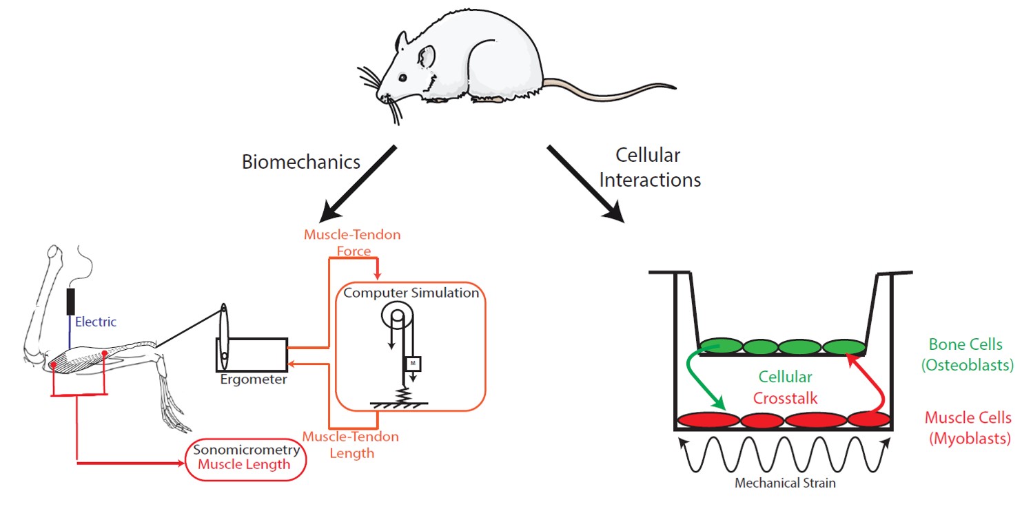
In collaboration with Dr. Gregory Sawicki, Mechanical Engineering, Georgia Institute of Technology
Musculoskeletal performance declines with aging, leading to conditions of sarcopenia and osteopenia, but underlying mechanisms for how they are related are poorly understood. We characterize tissue properties (in situ muscle stimulator, mechanical testing) and cellular interactions (co-culture) in a rat model to improve understanding of how age-related changes in musculoskeletal function may arise from cellular crosstalk.
Publications and Presentations:
- Doering J, Britt C, Cole J (2018) Understanding muscle-bone cellular crosstalk in aging. 8th World Congress of Biomechanics, Dublin, Ireland, Jul 8-12, Oral #O0172.
- Doering JA and Cole JH (2018) Optimizing a cell co-culture system to understand bone-muscle crosstalk. Orthopaedic Research Society Annual Meeting, New Orleans, LA, Mar 10-13, Poster #1549.
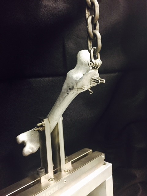
Testing Tension Band Wiring Configurations for Stable Bone Fracture Fixation
In collaboration with Dr. Simon Roe (Professor of Orthopaedics, College of Veterinary Medicine, NCSU)
Tension Band Wiring is used for surgical fixation of bone fractures in sites with large tensile forces, such as the greater trochanter (femur) or olecranon (ulna). This technique converts applied muscle/tendon/ligament tensile forces into compressive forces that minimize the fracture gap and promote healing and is commonly used in veterinary surgery. We are designing a better fixture system to isolate wire properties during mechanical testing and then testing different wiring configurations to determine an optimum one for trochanteric fracture fixation in veterinary patients. Our goal is to identify appropriate mechanical properties — such as yield point (defined by loss of initial wiring tension) and failure load — with cyclic loading that mimics clinical loading patterns. Comparing this information across wiring configurations will provide insight into the best techniques to use during treatment of veterinary fractures.
Publications and Presentations:
- Thompson E*, Robe RK*, Roe SC, Cole JH (2020). Influence of wire configuration on resistance to fragment distraction of tension bands placed in a greater trochanteric osteotomy model. Veterinary Surgery 49(4):710-718. DOI: 10.1111/vsu.13350. PMID: 31713901. * co-first authors
- Thompson E*, Roe SC, Robe AK, Cole JH (2016) Comparison of multiple tension band wire configurations on a greater trochanteric osteotomy model. American College of Veterinary Surgeons Surgery Summit, Seattle, WA, October 6-8. * 2nd Place, Small Animal Residents’ Forum Presentation, One AO Award
Testing Meniscus for Improved Tissue Engineering of Allograft Replacements
In collaboration with Drs. Jeffrey Spang (Orthopaedic Surgery, University of North Carolina), Elizabeth Loboa (Dean of Engineering, University of Missouri), and Matthew Fisher (Biomedical Engineering, UNC/NC State)
Meniscal tears are the most common knee injury. The meniscus does not repair well, and partial or complete surgical removal can be detrimental due to increased contact stresses. Current allograft replacements must be improved to mimic native tissue both biologically and mechanically. In collaboration with Dr. Elizabeth Loboa, we are measuring the material properties of various engineered tissues to determine the best replacement.
![]()
Image copyright eorthopod medical multimedia group, llc 2003: https://www.eorthopod.com/sites/default/files/images/knee_meniscus_transplant01_STILL.jpg
Publications and Presentations:
- Nordberg RC, Charoenpanich A, Vaughn CE, Fisher MB, Cole JH, Spang JT, Loboa EG (2016) A needle-punch method to enhance cellular infiltration of adipose stem cells in allograft menisci. Biomedical Engineering Society Annual Meeting, Minneapolis, MN, October 5-8, poster.
- Nordberg RC, Charoenpanich A, Vaughn CE, Griffith EH, Fisher MB, Cole JH, Spang JT, Loboa EG (2016). Enhanced cellular infiltration of human adipose derived stem cells in allograft menisci using a needle-punch method. J Orthop Surg Res 11(1):132. [DOI: 10.1186/s13018-016-0467-x]. PMCID: PMC5084349.